https://www.amazon.com/Why-We-Sleep-Unlocking-Dreams/dp/1501144316/ref=sr_1_1?ie=UTF8&qid=1510000478&sr=8-1&keywords=Why+We+Sleep
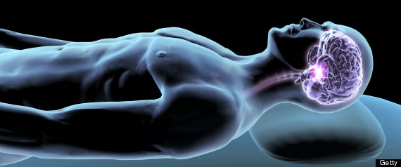
It's a testament to the occasional bone-headedness of scientists that sleep cycles were not discovered before the 1950s. You don't need an EEG recording to detect them. Simple observation of sleeper throughout the night will show you the main features. The most obvious of these is the rapid side-to-side eye movements that are easily seen even when the eyelids are closed (owing to the bulge of the cornea indenting the eyelid). Careful observation would reveal a host of other changes during REM sleep. These include an increase in breathing rate (as well as heart rate and blood pressure) and a sexual response (penile erection in men, erection of the nipples and clitoris together with vaginal lubrication in women). Even more striking are changes in muscle tone. The typical adult sleeper will change his or her position about 40 times a night without being conscious of this action. None of these motions, however, will occur during REM sleep. In REM sleep, there is no movement at all. In fact, there is not even any muscle tone: the body goes totally limp. It is almost impossible to have REM sleep in anything other than a horizontal position. Remember this the next time you are wrapped in an airline blanket and stuffed into your coach class seat like a scrofulous burrito for a trans-Atlantic flight: even if you manage to catch some sleep in your seat, you won't be able to enter REM sleep.

REM sleep is sometimes called "paradoxical sleep" because the EEG resembles that of the waking state, yet the subject is essentially paralyzed. The story here is that the motor centers of the brain are actively sending signals to the muscles but these signals are blocked at the level of the brainstem by inhibitory synaptic drive. This blockade affects only the outflow of motor commands down the spinal cord, not those of the cranial nerves that exit the brainstem directly to control eye and facial movements (as well as heart rate). Michel Jouvet of the University of Lyon showed that severing the inhibitory fibers that block motor outflow in cats resulted in a bizarre condition: during REM sleep the cats engaged in complex motor behaviors while keeping their eyes closed. They ran, pounced, and even seemed to eat their imagined prey. Although we can't know this for certain, they appeared to be acting out their dreams...A similar phenomenon is seen in a human condition called REM sleep behavior disorder, which mostly affects men over 50. This disease causes dream-enacting behaviors during the REM period of sleep, including kicking, punching, jumping, or even running. Not surprisingly, these violent behaviors can often result in injury to the patient or to his or her bedmate. In most cases, this disorder is successfully treated by a bedtime dose of the drug clonezepam...which works by boosting the strength of synapses that use the inhibitory neurotransmitter GABA. REM sleep behavior disorder is different from conventional sleepwalking, which occurs only during non-REM sleep.
Humans show changes in sleep over the life cycle, with the proportion of the time spent in REM sleep decreasing from about 50 percent at birth to 25 percent in mid-life and 15 percent among the elderly (a decrease in REM is also seen over the lifespans of cats, dogs, and rats). If we compare our sleep with that of other mammals, we find that we are more or less in the center of the range bounded by the duck-billed platypus, which spends about 60 percent of its sleeping life in REM, and the bottlenose dolphin, which has a REM proportion of only 2 percent. There is no obvious relationship between degree of REM sleep and brain size or structure across mammalian species...Non-REM sleep appears to have evolved as early as the fly (about 500 million years ago), but true REM sleep is found only in warm-blooded species. It is present in the most primitive surviving mammals (such as the platypus and the echinda) as well as in birds, but appears to absent in reptiles and amphibians.
So, with knowledge of sleep cycles, we can return to our main question "Why is sleep necessary?" with a bit more sophistication. Really, two separate questions are warranted: What are the key functions of sleep composed of...And, what are the key functions of cycling sleep in which REM and non-REM periods alternate, as is found in mammals and birds? It may be that the previously mentioned ideas that sleep is required for restorative functions, energy conservation, and maximizing feeding efficiency while minimizing danger from predation are appropriate for non-REM sleep alone. Cycling sleep is serving some function that only emerges in mammals and birds and that is most important in early life...
...
One interesting study of human learning and sleep deprivation comes from the laboratory of Jan Born at the university of Lubeck in Germany, where investigators sought to test the notion that a night's sleep can help yield insight into a previously intractable problem. To do this, a numerical problem was devised that could be solved by sequential application of simple rules. The experimenters embedded within the problem a shortcut that, if appreciated, could allow the subject to respond much more quickly than through the sequential-application method...None of the participant's recognized the shortcut in the first block of trials. After a night's sleep, though, 13 of 22 subjects had the insight to recognize the shortcut, while, in a different group of subjects, who were not allowed to sleep over a similar interval, only 5 of 22 found the shortcut. The experimenters' conclusion: sleep inspires insight.
https://www.nytimes.com/2017/02/02/science/sleep-memory-brain-forgetting.html?_r=0
WHEN YOU SLEEP YOU ERASE MEMORIES TO CREATE ROOM TO MAKE NEW MEMORIES.
WHEN YOU SLEEP YOU ERASE MEMORIES TO CREATE ROOM TO MAKE NEW MEMORIES.
A large number of studies have sought to interfere with REM sleep by waking humans or lab animals when an EEG recording indicates that they have entered a REM stage. Selective REM deprivation has been reported to interfere with memory consolidation for a number of learning tasks. In some cases the results have been dramatic: in one report, when humans were trained in a visual texture discrimination task, in which reaction time is taken as a measure of learning, they showed no evidence of learning after either a normal sleep or sleep in which non-REM periods were selectively disturbed. It's important to note that REM deprivation seems to interfere specifically with the consolidation of memories for rules, skills, procedures, and subconscious associations (nondeclarative memory) but not memories of facts and events (declarative memory). Thus the people who spent a REM-deprived night following visual texture discrimination training still had clear memories of the training session (an event) but did not retain their quick reaction times in the task (a nondeclarative skill).
The timing of REM sleep also appears to be important. REM sleep must occur within 24 hours of the training experience in order for it to improve memory consolidation. People who learn a new skill or procedure during the day and then miss that night's sleep will not show any improvement following sleep on the second night. A similar effect is seen in rats, but the interval is reduced: REM sleep must occur within 4-8 hours of training to have a beneficial effect.
REM sleep also appears to be associated with "playback" of the previous day's memories. Kendall Louie and Matt Wilson of MIT used arrays of electrodes to simultaneously record from large numbers of "place cells" in the hippocampus of rats as they repeatedly ran a unidirectional path in a circular track to obtain a food reward. The experimenters were able to see sequential activation of place cells coding for various locations on the circular track as the animal ran. Then recordings were continued as the animal slept after training. Amazingly, these same patterns of hippocampal place cell activation were replayed during REM sleep. The replay wasn't a perfect spike-for-spike reproduction of the waking activity. Sometimes the pattern was a bit degraded and sometimes the pattern was recognizable from the waking experience, but the overall speed of the activity had changed. Nonetheless, this study and several others like it from different laboratories, have found statistically significant reactivation of neuronal ensemble activity during REM sleep following training....
11:30
.jpg) |
| 3 A.M. |
With each bit of information we process, millions of neurons are active. The problem is, much as a running motor generates exhaust fumes, all of this neural activity leads to a serious buildup of cellular waste products. Eventually our cells need a nap. Neuron "exhaust" takes the form of molecules, including one called adenosine. Prompted by the adenosine buildup, our bodies nudge us into bed when our batteries need recharging.
Anyone who has struggled to stay awake while driving knows the relentless pressure adenosine exerts on us to stop and sleep. Adenosine itself doesn't cause the sleepiness; it's a messenger that simply signals the surrounding cells to settle down. Caffeine blocks this sleepy message. Here's how.
Our brain cells communicate by passing chemical messages like adenosine to one another. Messenger molecules are teamed up with specific listeners - called receptors - on other cells. Adenosine and its receptors are exquisitely matched, like tiny locks that open only with the proper miniature key. When a cell releases adenosine, it fills adenosine receptors on nearby cells, passing along the message to sleep.
And the production of adenosine continues throughout our day, more and more receptors are filled. Increasingly our brain cells become sluggish, regardless of how strongly they are stimulated. We become tired. As we sleep, the night shift sweeps the adenosine away. When we awake, we feel better because we literally are more clear-headed.
But let's say we don't have the luxury of climbing into bed when we feel tired. Instead, we reach for a soda or a double espresso. The caffeine we ingest makes a beeline for our brains, and once there, it bubbles around between the cells. Here, because of a chance similarity in shape, the caffeine slips into some of those receptors intended for adenosine.
Once nestled in these slots, caffeine just camps out, blocking adenosine from doing its job. So perhaps we've been up for hours, working like crazy, and our brains are awash in adenosine. We should be dead tired. But with many of the receptors blocked by caffeine, the adenosine can't pass on the message that we ought to go to bed. Instead we feel surprisingly alert and still ready to take on the world.
Caffeine keeps us awake because it interrupts the normal sleep signaling system. Some drugs obstruct the body's natural signaling system while others amplify messages. In every case, however, drugs masquerade as naturally occurring compounds, tricking our brains...
Mean Genes: From Sex to Money to Food Taming Our Primal Instincts. Burnham, Phelan, 62-63.

.jpg)
.jpg)

.jpg)
.jpg)
.jpg)
.jpg)
.jpg)

